Today at the microscope: Diatom and Coccolithophore BFFs

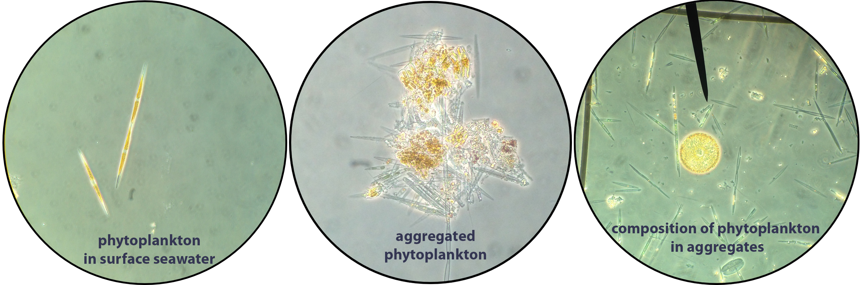
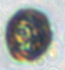
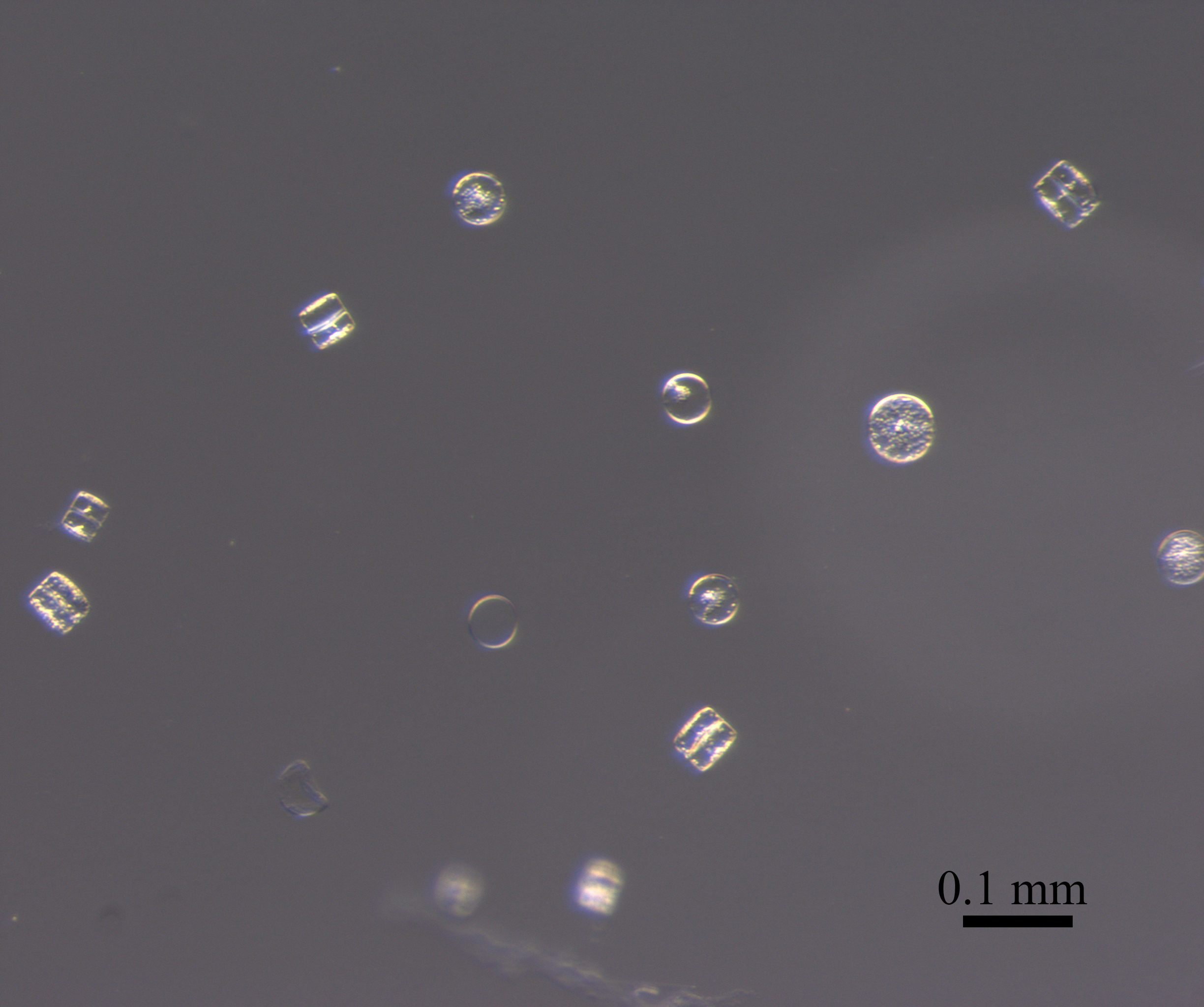
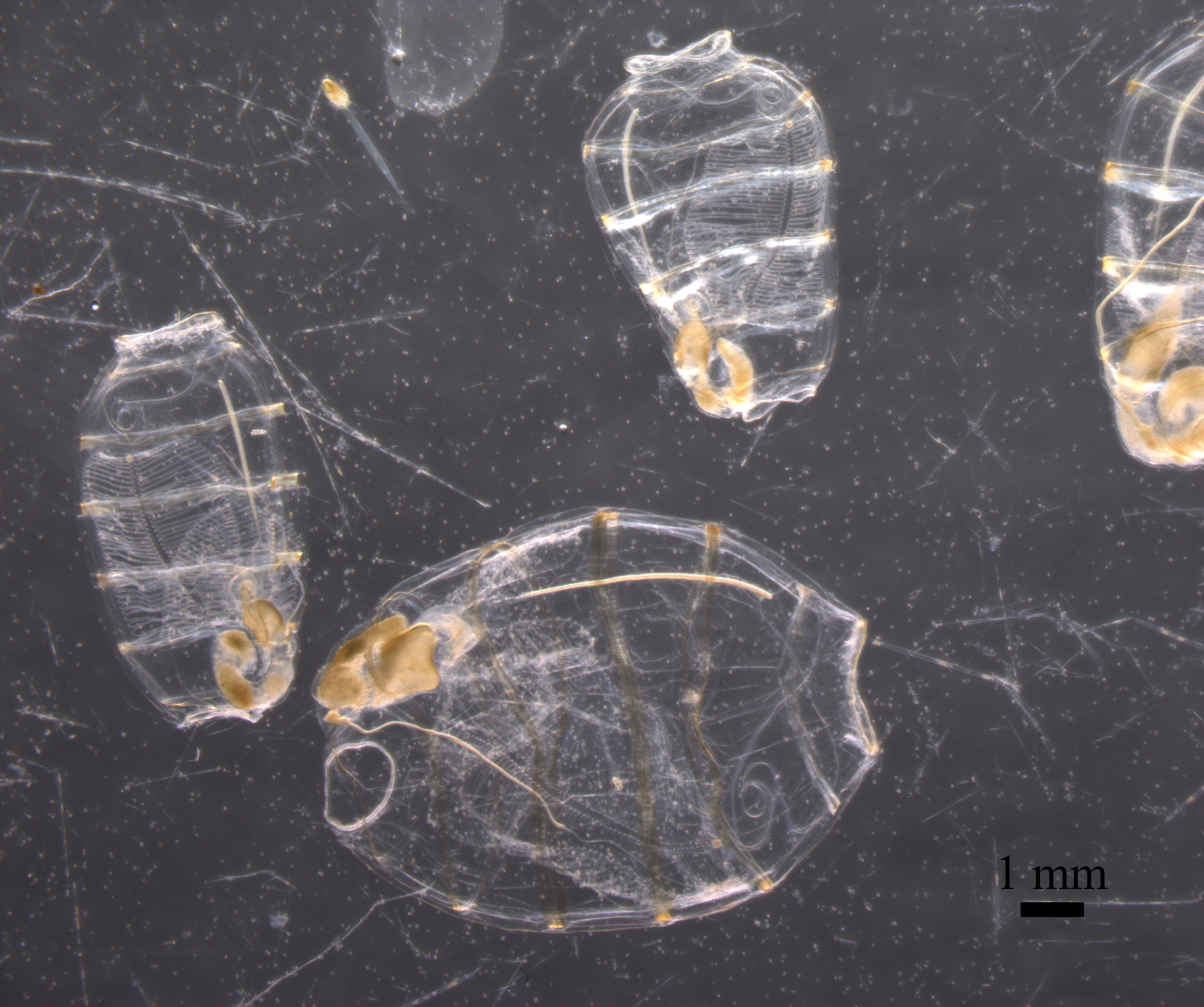
Yesterday as I was looking under the microscope at particles collected from the bottom of the ocean, and I came across this beautiful cell(s):


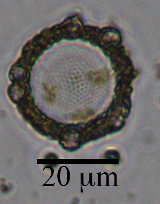
This centric diatom is surrounded by a halo of smaller coccolithophore cells.  The circle in the center is the diatom.  You can see the nano-patterns on the silica valve and some yellow/brown-ish chlorophyll inside the cell.  Surrounding the cell are atleast 6 coccolithophores cells (smaller circles) and a ring of their coccoliths (the brown-ish looking mass).  The silica frustules of diatoms and calcium carbonate coccoliths of coccolithophores are relatively heavy biominerals, and may increase carbon export out of the surface ocean by “ballasting” sinking particles.  There is some debate in the literature about which biomineral is more important in exporting carbon.  This diatom-coccolithophore association illustrates my opinion: they are probably both very important.

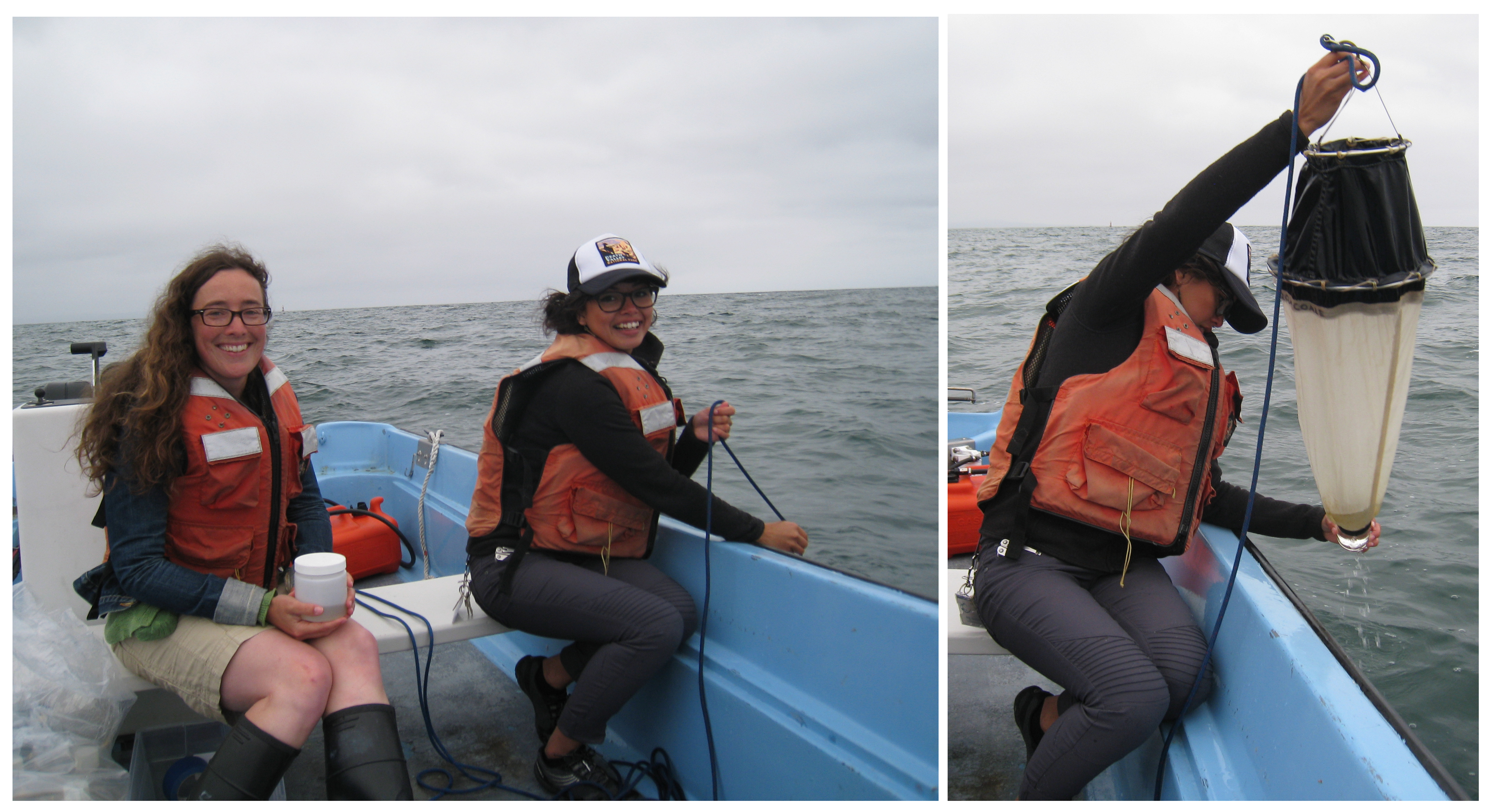
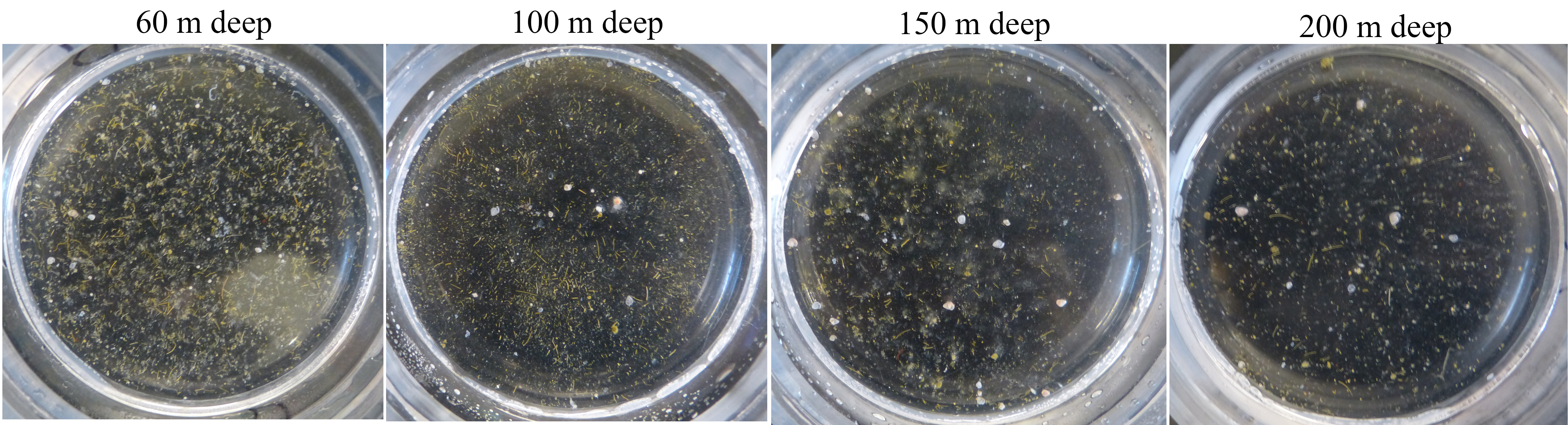
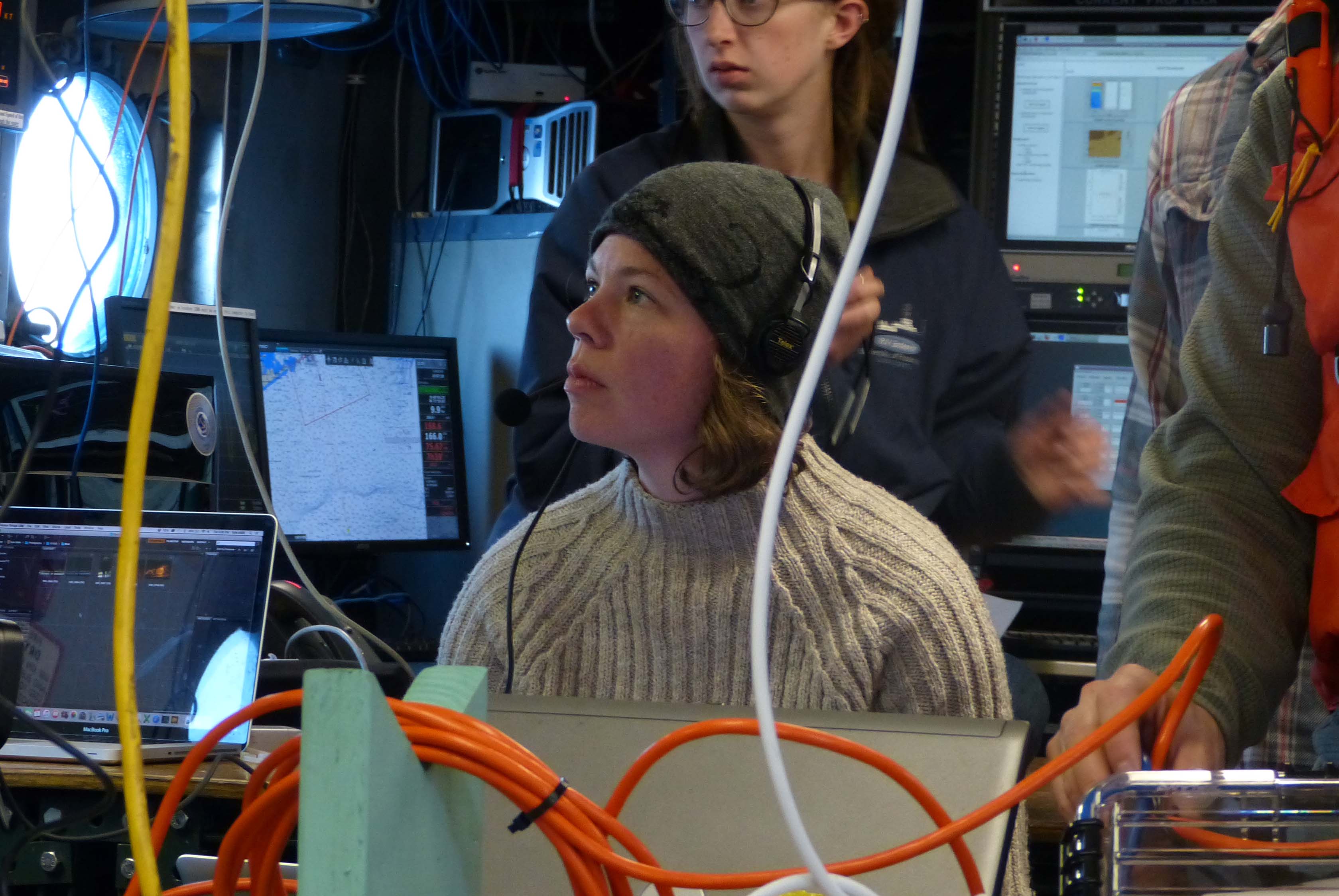
This sample was collected in a sediment trap 4000 m deep, at the Station M time-series.  I am currently counting phytoplankton within the particles collected over the past 30 years in these sediment traps in collaboration with Ken Smith and Crissy Huffard at MBARI, and thanks to support from  a 2017 New Faculty Award from the California Sea Grant.

Read more from Sea Grant here:
https://caseagrant.ucsd.edu/news/special-focus-awards-support-new-faculty-resilient-coastal-communities
https://caseagrant.ucsd.edu/project/how-does-climate-change-affect-the-export-of-phytoplankton-to-the-seafloor

New microscope, ready to bring out to sea!

This week I received my lab’s first microscope!  It is an Olympus SZX16 stereo microscope. Stereo Microscope
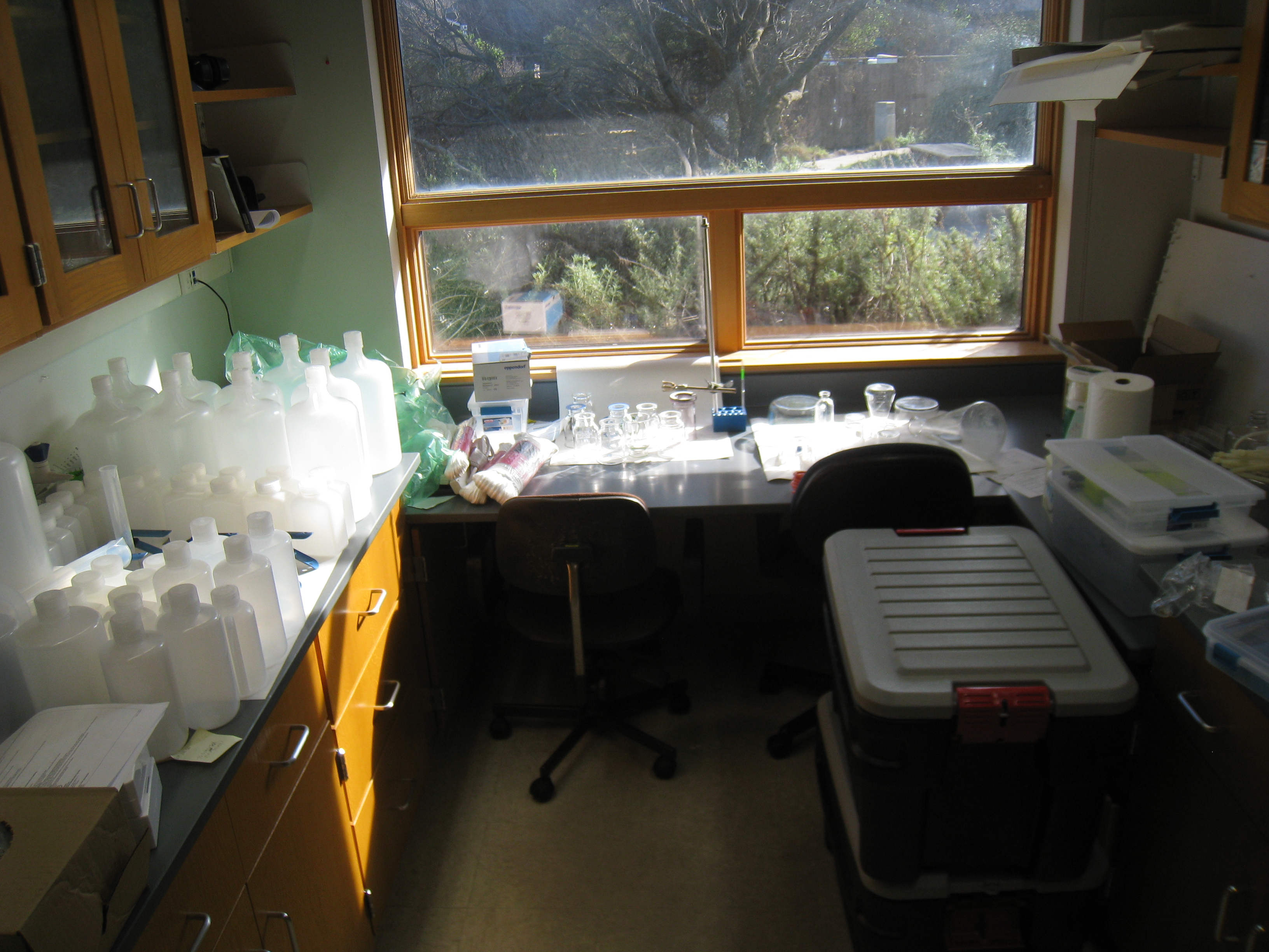
This microscope will be used to image and quantify sinking particles collected in sediment traps.  It also fits easily into a carry-on size bag so that I can safely transport it out to sea.

img_0002


This microscope is coming with me on the upcoming Sea2Space cruise aboard the R/V Falkor (leaving next week!), and is part of a new research project recently funded by NSF.  I received it just in time to test out its imaging capabilities with a preserved plankton net tow I collected last summer in Monterey Bay.  This sample contained a type of gelatinous zooplankton called doliolids and a bloom of diatoms.  I love how we can see both large and small things:

The doliolids are between 0.5 cm and 1 cm long, while the diatoms are about 0.005 cm long (or ~50 micrometers).  The diatoms in this image are called Coscinodiscus.

diatoms doliolids

I can’t wait to image sinking particles next month with my first microscope, and I’m already dreaming about microscope #2 for the lab.

New NSF award to link plankton with sinking particles

A new project in my lab was recently funded by the National Science foundation. (My first NSF award!) The title of the project is:

“Collaborative Research: EAGER: Particle-specific DNA sequencing to directly observe ecological mechanisms of the biological pump”

This project is a collaboration with my co-principal investigators Margaret Estapa (Skidmore College) and Melissa Omand (University of Rhode Island) and will fund work during a month-long research cruise later this winter.

drill-and-micropippeter
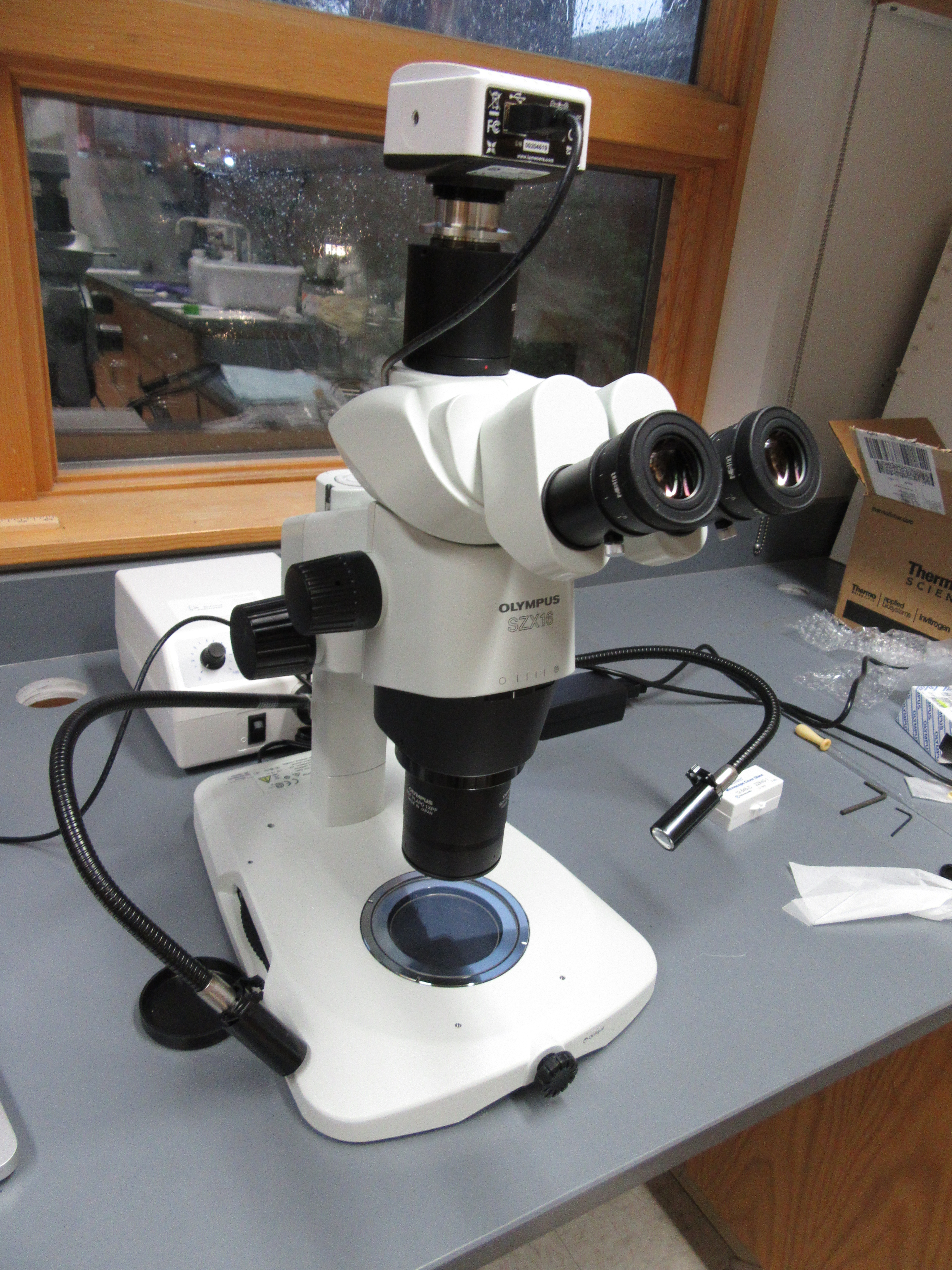
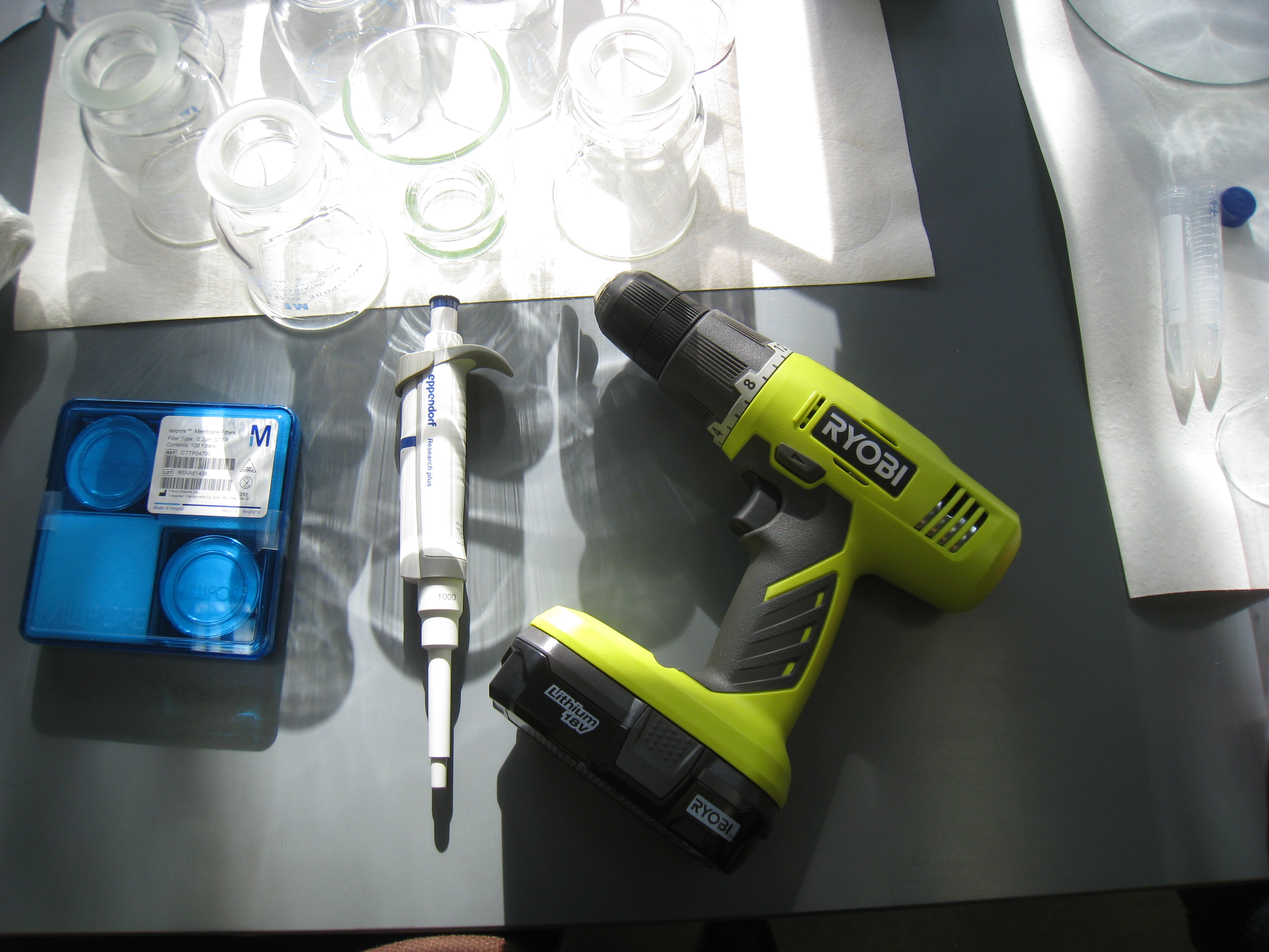
Like any good oceanographer, I immediately celebrated by purchasing a drill and a micropippetor.

Here is the non-technical abstract of the project:
Carbon is fixed into organic matter by phytoplankton growing in the surface ocean, and is naturally sequestered in the ocean interior when particles and organisms sink: a process called the “biological pump”. Because of its recognized influence on the global carbon cycle, ocean scientists have studied the biological pump for decades. However, we still do not have a sufficient understanding of the underlying processes to accurately quantify and predict carbon cycling. Much of this uncertainty stems from an inability to directly link specific plankton in the surface ocean with the types of particles sinking out of the surface ocean. To address this missing link in biological pump research, this work will directly observe how plankton are transported out of the surface ocean using novel, particle-specific observational approaches embedded within an interdisciplinary field program that will finely resolve upper ocean plankton groups and the resulting amount of sinking carbon across space and in time. The genetic identity of organisms within different types of sinking particles will be determined by sequencing the genetic contents of individually collected particles. This new application of a molecular method will definitively link surface plankton with sinking particles at 5 locations across the Pacific Ocean. This work has the potential to transform our understanding of the biological pump by identifying previously unknown links between surface ecosystems and sinking carbon particles. Because this work is embedded within an interdisciplinary field program, including biogeochemical modelers and remote sensing scientists, these data will feed directly into new models of the biological pump, improving our ability to quantify and predict carbon uptake by the ocean.

This project was funded by the National Science Foundation Biological Oceanography program through their EAGER funding opportunity (Early-concept Grants for Exploratory Research).

We will be sailing on the Schmidt Ocean Institute’s vessel, the R/V Falkor, from Hawaii to Seattle.  Along the way we will stop and deploy a variety of sediment traps and sensors to measure and collect sinking particles.  This cruise is lead by chief scientist Ivona Cetinic (NASA) and will include an exciting interdisciplinary team of scientists including optical oceanographers, remote sensing scientists, and physical- chemical- and biological- oceanographers.  The cruise has a website and is named the “Sea to Sky” cruise.  If you want to check out the ship, of course the Schmidt Ocean Institute has a google-street-view-like virtual tour online.  Here is the lab space we’ll be working in.

I am super excited about this project!  Stay tuned for more updates as it progresses.

Cruise preparations have begun.


Diatom community composition and aggregation

Posted by CSUMB REU student Melia Paguirigan:

This summer I participated in the California State University Monterey Bay Research Experience Undergraduate program, with Dr. Colleen Durkin as my mentor. Our project investigated the role of diatom community composition and morphology in aggregation. We collected whole seawater samples from Monterey Bay.

Durkin_Paguirigan_Whalercollection
collecting seawater and a plankton net tow on an MLML boston whaler offshore of Moss Landing

Then I used a roller table to make aggregates.

In the lab, we used microscopy techniques to quantify the community composition of the aggregates and the corresponding surface water.

Aggregation_experiment_REU


The data was then used to identify if diatoms differentially aggregate and if morphology was driving differential aggregation. Throughout this process I was able to become familiar with over 20 diatom genera, using their shape and colony formation as identifiers.

(Note from Colleen: Melia estimates that she counted >21,000 individual diatom cells this summer!  She found significantly different phytoplankton compositions in aggregates compared to the total community in seawater, suggesting that some genera tend to be incorporated into aggregates more than others.  Melia plans to present this data at a conference later this year.)


Early summer particle flux at the New England continental shelf break

Last week I visited Melissa Omand’s lab at the University of Rhode Island to analyze sediment trap samples collected on the R/V Endeavor.  Unfortunately I was not able to go on the cruise, but I was still lucky enough to look at the exciting samples they brought back.

June_geltrap_jars

The types of samples collected on this cruise were very similar to those we collected in November.  Sediment traps collected sinking particles in the water column.  Above you can see how the number of sinking particles decreased with depth. These are images of the polyacrylamide gel jars placed in the bottom of traps at 4 depths spanning the upper mesopelagic zone.  The 60 meter trap was full of zooplankton fecal pellets (the long stringy particles).  At 150 meters, fluffy diatom aggregates appeared (mostly containing Pseudo-nitzschia), but sinking fecal pellets were also still abundant.  At high magnification, it became apparent that the traps were also chock-full of tiny little particle which turned out to be individual sinking coccolithophore cells. They are only about 10 micrometers big, and difficult to image clearly, but you can just make out the circular coccolith plates covering the outside of this cell .

Cocco

Although there is still much analysis to be done, these first observations are already exciting and represent a very different particle export environment than what we observed last November, which was dominated by large organic aggregates and large diatom cells.

On a related note, the URI Inner Space Center created several short videos about the research cruise last November.  Here are the films explaining the sediment traps.

Meg Estapa explaining the neutrally buoyant sediment traps:

Me, explaining the particle work:


Pat Kelly explaining the surface-tethered sediment trap array and in-situ pumps:

New publication: sinking phytoplankton associated with carbon flux

Our study of phytoplankton associated with sinking particles was recently published in the journal Limnology and Oceanography. (link to open access publication)

The amount of carbon that sinks out of the surface ocean in the form of organic particles is highly variable and difficult to predict, in part because the ecological processes that lead to the export of these particles are complicated.  In this study, we attempted to resolve the mechanism of particle export across a large ocean basin, with a special focus on phytoplankton cells.

TDurkin et al 2016 Figure 3his data was collected in 2013 during a 45 day research cruise on the R/V Knorr (aka: the DeepDOM cruise).  We sailed from Uruguay to Barbados and deployed sediment traps all along the way (see locations on the map).  You can see a video about this project here, and on the “Science Videos” page.

I used sediment traps containing a gel layer at the bottom to resolve the identity of individual particles and cells sinking out of the surface ocean (see micrographs in the map figure).  The observation that excited me the most was the presence of individual phytoplankton cells in the gel layer.  How could such tiny “particles” sink out of the surface ocean?  We used a statistical approach to hypothesize how these tiny, slow-sinking cells  were transported out of the sunlit surface ocean where they grow.

Durkin et al 2016 Figure 8

At most locations, phytoplankton appeared to be associated with sinking fecal pellets and aggregates; they were most likely carried along with these fast sinking detrital particles.  However, at several locations sinking phytoplankton cells did not seem to be associated with detrital particles.  At two locations, intact and living diatoms appeared to be sinking by themselves, suggesting that they could sink fast enough to be transported out of the surface ocean.  Alternatively, they may have been transported by physical mixing.  Coccolithophores were particularly dominant at 3 of the observed locations, and we hypothesize that they were either transported by sinking, physical mixing, or in association with detrital aggregates.

We hope that these methods will continue to resolve the mechanisms of “the biological pump” in future studies.  With a better understanding of the types of particles responsible for carbon uptake by the ocean, and mechanisms responsible for their transport to the deep sea, the global carbon cycle can be better quantified.

This project was a collaboration with Ben Van Mooy (WHOI), Sonya Dyhrman (Columbia/LDEO), and Ken Buesseler (WHOI), who are also coauthors.

Surface phytoplankton and sinking particles offshore of Rhode Island

The “EN572” cruise aboard the R/V Endeavor was a success.  My goals on this cruise were to link the phytoplankton communities growing in the surface with the particle types sinking out of the surface.  We observed a very abundant and diverse phytoplankton community, including many different species of diatom and dinoflagellates.

Phytoplankton observed on EN572 cruise

The sinking particles were dominated by large, fluffy aggregates.  The aggregates contained many of the phytoplankton cells that we observed in the surface waters.

Aggregate containing diatoms

Now that we are all back on land, it is time to analyze the samples and all the data that we collected.  Stay tuned!

Departure and telepresence

This morning we set sail, heading for the Rhode Island continental shelf break.  We are sailing on the R/V Endeavor:

R/V Endeavor
R/V Endeavor

Unfortunately, Melissa, the chief scientist had to stay on the dock, because she is beyond the pregnancy threshold for sailing on a cruise.

Melissa had to stay behind.

Instead, Melissa will be acting as chief scientist from shore and will stay connected with the ship through the University of Rhode Island’s “telepresence” capabilities.  Camera and microphones connect Melissa on shore with the ship.  Meg Estapa is acting as the on-ship chief scientist.

Meg coordinating the sailing plan with Melissa over the video connection.

Video from our cruise is streaming live on Youtube, and at this link:

http://innerspacecenter.org/en572